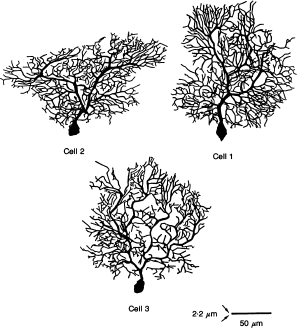
| Figure 1: | Reconstruction of HRP-labelled Purkinje cells from
guinea-pig cerebellum. Three Purkinje cells were reconstructed in 3D,
using a semi-automatic computerized tracing system (Eutectic Inc). Cells
were represented by 1400–2100 sample points (each point specified by x,
y and z coordinates and the diameter of the structure at that point) that
were later used to build the corresponding cable and compartmental models.
Dendritic spines were not included in this reconstruction but were added to
the models assuming that dendritic branches with diameters < 2.2μm bear
10 spines (um length)-1, each spine having an area of 1μm2. The length of
the scale bar represents 50 μm of dendritic length whereas the thickness of
the scale bar represents 2.2 μm dendritic diameter. |